
Cologuard®
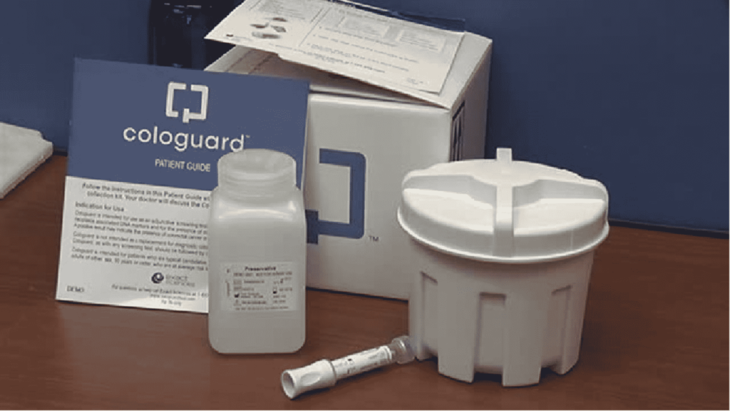
Cologuard® Non-Invasive Colorectal Cancer Screening Option Colorectal cancer is a prevalent health concern, but with advancements in medical technology, there are now more accessible and non-invasive screening options available. One such option gaining attention is Cologuard. In this section, we delve into the specifics of Cologuard and its significance in colorectal cancer detection. The Science Behind Cologuard Cologuard is a molecular stool test designed to detect both colorectal cancer and advanced adenomas. It employs advanced DNA analysis and fecal immunochemical testing to identify altered DNA and blood in the stool. Understanding the scientific basis of Cologuard is crucial for patients seeking an effective and convenient screening method. Cologuard vs. Colonoscopy While both Cologuard and colonoscopy serve as screening tools, they adopt distinct approaches and provide different information. Cologuard detects proteins shed from colon cancer and polyps, while colonoscopy visually identifies the polyps themselves. Colonoscopy, despite its effectiveness, carries a low risk of complications, including reactions to sedatives or possible puncturing of the bowel. Cologuard, in contrast, eliminates these risks. However, a 2019 study identified limitations, including the potential for false negatives, overlooking larger polyps, and a higher risk of false positives compared to colonoscopies. Notably, Cologuard and colonoscopy can complement each other in a screening regimen. Cologuard can serve as a non-invasive, first-line test for individuals at average risk, providing an additional layer of early detection. Eligibility for Cologuard The target demographic for the Cologuard test includes individuals at average risk, particularly those between 50 and 75 years old. Screening recommendations from the U.S. Preventive Services Task Force (USPSTF) suggest initiation at age 50, while the American Cancer Society recommends screening from age 45. For individuals with an increased risk of colon cancer due to family history, inherited mutations, or other known risk factors, consultations with healthcare providers are vital. These discussions should explore the possibility of commencing screening even earlier. Interpreting Cologuard Test Results Following the submission of a stool sample, Cologuard test results, delivered as either “negative” or “positive,” are sent to the attending physician. Negative results indicate the absence of atypical DNA or hemoglobin biomarkers, suggesting no signs of colon cancer or precancerous polyps. In the event of a positive result, further testing, typically a colonoscopy, is recommended to confirm and address potential issues. It is crucial to acknowledge the presence of false positives and false negatives in Cologuard results. A 2014 clinical study revealed a 13 percent false positive rate and an 8 percent false negative rate, highlighting the importance of cautious interpretation. In the ever-evolving landscape of colorectal health, Cologuard stands as a valuable addition to our arsenal of screening tools. Its non-invasive nature and accessibility contribute to increased screening compliance, potentially leading to earlier detection and intervention. However, maintaining a nuanced understanding of its benefits and limitations is paramount. back to conditions page Facebook-f Youtube Linkedin-in Instagram
Small Bowel Capsule Enteroscopy
SBCE SMALL BOWEL CAPSULE ENTEROSCOPY A novel diagnostic tool that allows direct visualization of the small intestine. A vitamin-sized capsule containing a miniature camera is swallowed by the patient and takes thousands of pictures as it travels through the gastrointestinal tract. Overview of Small Bowel Capsule Enteroscopy Small bowel capsule enteroscopy was approved by the FDA in 2001 as a tool to examine the small intestine, which previously could not be visualized well with traditional endoscopy. The capsule camera takes approximately 50,000 images during its travels through the GI tract, providing significantly more visualization than other methods. Capsule endoscopy is a non-invasive way to diagnose small bowel abnormalities and diseases, such as Crohn’s disease, ulcers, celiac disease, small bowel tumors, and sources of GI bleeding. Procedure Patients fast overnight before capsule endoscopy. The next morning, they ingest the vitamin-sized capsule with water. Sensors are placed on the patient’s abdomen with adhesive pads, and are connected to a data recorder worn in a belt around their waist. Patients go about normal daily activities as the capsule camera takes images, while the sensors track its location and collect the images. After 8-12 hours, the sensors and recorder are removed, and the images are downloaded to a computer workstation for physician review. Patients pass the capsule naturally in their stool within 1-2 days. Interpretation of Results A physician carefully reviews the thousands of capsule images looking for abnormalities. Diagnostic findings include ulcers, bleeding, tumors, blockages, polyps, villous atrophy from celiac disease, and signs of Crohn’s disease. The images provide direct visualization to confirm diagnoses. However, capsule location can only be estimated based on image transit times. If an abnormality is seen, its exact location may be unclear. Another endoscopic procedure may be required to take biopsies or provide therapy. Limitations A major limitation of capsule endoscopy is the lack of therapeutic capabilities. It is strictly a diagnostic tool. Additionally, there is a small risk of capsule retention if a stricture is encountered. Capsule retention requires surgical or endoscopic removal. Some patients may have difficulty swallowing the capsule. Improper sensor placement or interference can also cause poor image quality. However, capsule endoscopy is considered very safe with few complications. Small bowel capsule endoscopy allows detailed visualization of the small bowel mucosa. It is a minimally invasive and safe procedure to identify causes of GI bleeding, inflammation, tumors, celiac disease, and other small bowel abnormalities. Despite some limitations, capsule endoscopy is a revolutionary way to diagnose diseases of the previously hard-to-reach small intestine. back to conditions page Facebook-f Youtube Linkedin-in Instagram
Esophageal Dilation
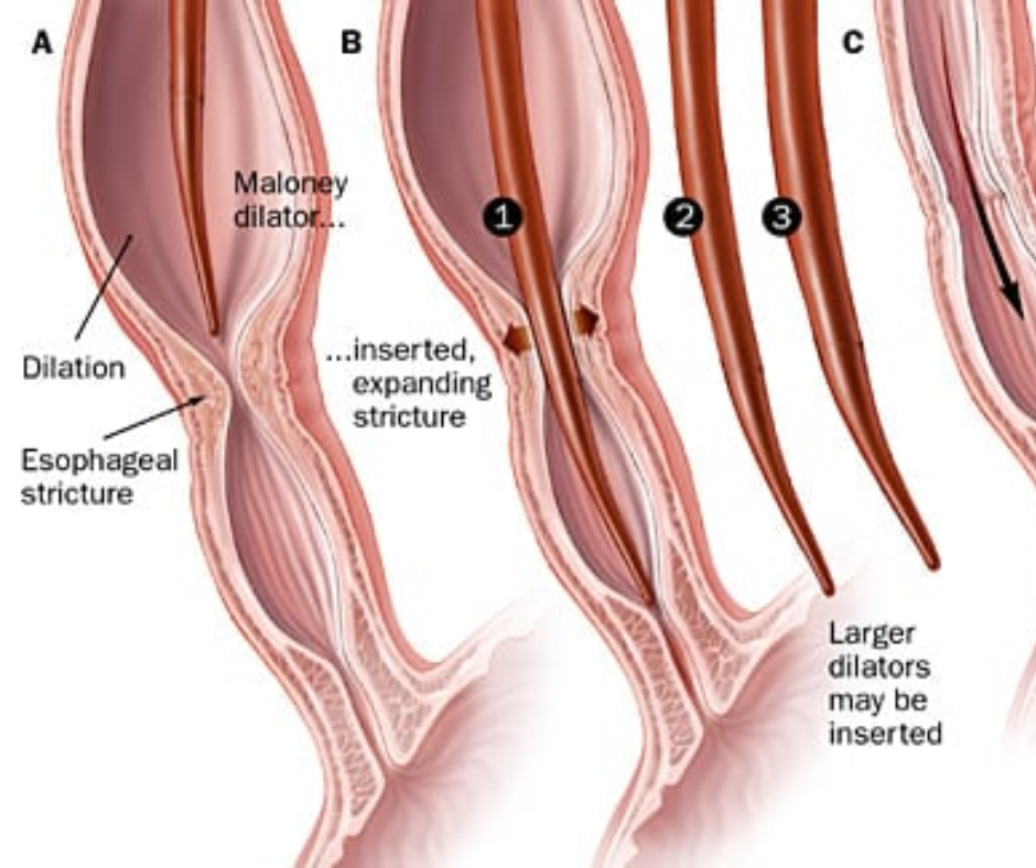
Esophageal Dilation Introduction to Esophageal Dilation Esophageal dilation is a medical procedure that aims to address difficulties in swallowing and other esophageal issues. This therapeutic intervention involves the gentle stretching of the esophagus to alleviate narrow passages and promote better functionality. Indications for Esophageal Dilation Esophageal dilation is commonly recommended for patients experiencing dysphagia, a condition marked by difficulty swallowing. Other indications include the presence of esophageal strictures, rings, or narrowing, which can impede the smooth passage of food and liquids. Understanding these indications is crucial for patients to grasp why their healthcare provider may recommend this procedure. The Procedure During esophageal dilation, a specialized dilator is carefully introduced into the esophagus under endoscopic guidance. The dilator is then gently inflated, causing controlled stretching of the narrowed areas. This procedure is typically performed on an outpatient basis, with patients returning home on the same day. A comprehensive understanding of the process empowers patients to approach the procedure with a sense of preparedness. Preparing for Esophageal Dilation Effective preparation is paramount to ensure the success and safety of the esophageal dilation procedure. Patients are advised to fast for a specified duration before the procedure to minimize the risk of complications. Additionally, it is crucial to inform the healthcare team about any medications, allergies, or existing medical conditions. Adhering to these preparatory measures contributes to a smoother and more efficient dilation process. Potential Risks and Aftercare While esophageal dilation is generally considered safe, it is essential for patients to be aware of potential risks and complications. These may include mild discomfort, bleeding, or perforation of the esophagus in rare cases. Following the procedure, patients may experience a sore throat or mild chest discomfort, which typically resolves within a short period. Understanding these aspects aids patients in recognizing normal post-procedural sensations versus signs that warrant medical attention. In conclusion, esophageal dilation is a well-established procedure designed to enhance the quality of life for individuals grappling with esophageal issues. This guide offers valuable insights into the purpose of the procedure, its indications, the actual process, and crucial steps for preparation and aftercare. Armed with this knowledge, patients can approach esophageal dilation with confidence, actively participating in their healthcare journey. back to conditions page Facebook-f Youtube Linkedin-in Instagram
Esophageal Manometry
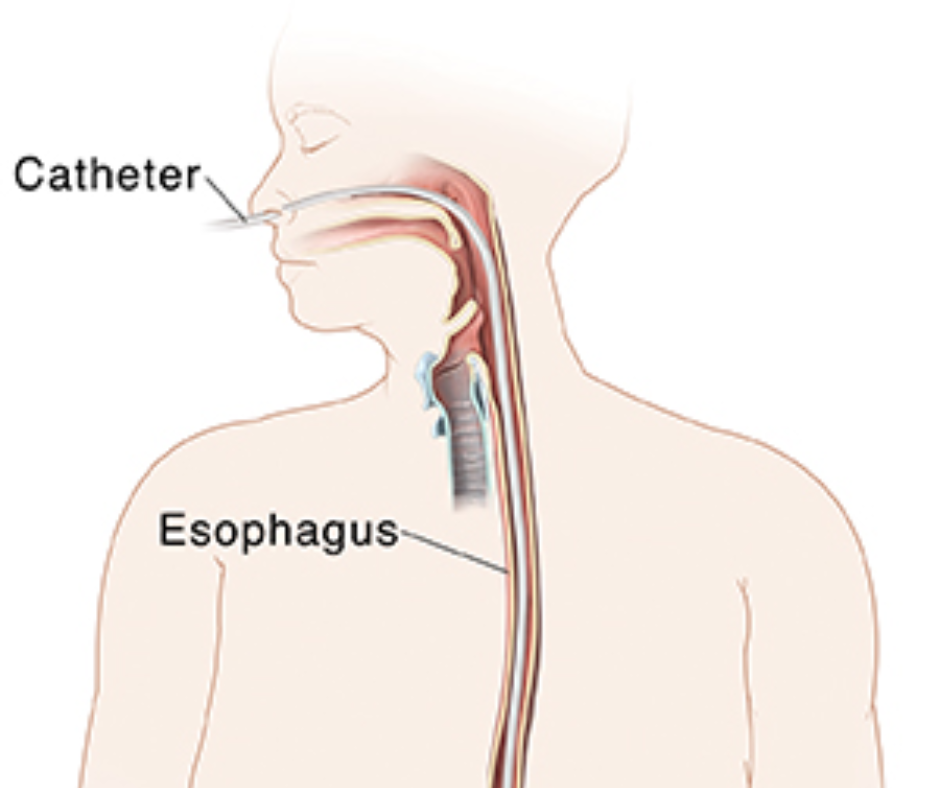
Esophageal Manometry Understanding Esophageal Manometry Esophageal manometry is a diagnostic procedure employed by gastroenterologists to assess the function of the esophagus and lower esophageal sphincter (LES). This test aids in identifying various esophageal motility disorders, helping healthcare professionals tailor effective treatment plans. Purpose of Esophageal Manometry Esophageal manometry serves the primary purpose of evaluating the muscular contractions and coordination within the esophagus. By measuring the pressure and motility of the esophageal muscles, this procedure assists in diagnosing conditions such as achalasia, gastroesophageal reflux disease (GERD), and dysphagia. Patient Preparation for Esophageal Manometry Before undergoing esophageal manometry, patients need to follow specific guidelines to ensure accurate and reliable results. Preparation often includes fasting for a specified period, discontinuation of certain medications, and avoiding foods that may affect test outcomes. Your healthcare provider will provide detailed instructions tailored to your individual health needs. The Procedure: What to Expect During esophageal manometry, a thin, flexible tube equipped with pressure sensors is passed through the nose and into the esophagus. Patients may experience a mild discomfort or urge to swallow as the tube is positioned. The procedure typically takes around 30 to 45 minutes, during which time you will be asked to swallow water or a specialized solution to assess esophageal function. Post-Procedure Considerations Following the completion of esophageal manometry, most patients can resume their regular activities. Some individuals may experience mild throat irritation or discomfort, but these symptoms are generally short-lived. Your healthcare provider will discuss the test results with you and recommend appropriate steps based on the findings. In conclusion, esophageal manometry is a valuable diagnostic tool that provides crucial insights into esophageal function. By understanding the purpose of the procedure and adequately preparing for it, patients can contribute to the accuracy of results and facilitate the development of effective treatment strategies for their specific esophageal conditions. If you have any concerns or questions about esophageal manometry, do not hesitate to discuss them with a healthcare provider. back to conditions page Facebook-f Youtube Linkedin-in Instagram
Upper Endoscopy/EGD

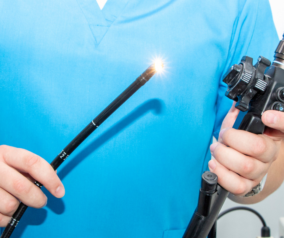
Upper Endoscopy/ EGD Upper GI Endoscopy (EGD) Upper gastrointestinal endoscopy, or EGD, is a diagnostic procedure used by healthcare professionals to investigate symptoms like heartburn or abdominal pain. Also known as esophagogastroduodenoscopy, this overview covers its purpose, conditions diagnosed, procedural aspects, and possible alternatives. Exploring the Upper Digestive Tract An EGD utilizes an endoscope, a flexible tube equipped with a camera and light, to meticulously examine the upper digestive tract. This includes the esophagus, stomach, and the upper part of the small intestine known as the duodenum. Through this procedure, physicians obtain visual insights into the digestive tract, capturing detailed images and, if necessary, conducting biopsies of abnormal tissue. Indications for an EGD The necessity for an EGD arises in cases where patients exhibit symptoms indicative of upper digestive tract disorders. These may include bleeding, heartburn, nausea, vomiting, abdominal pain, trouble swallowing, unexplained weight loss, and more. Conditions diagnosed through EGD encompass bacterial infections, celiac disease, Crohn’s disease, GERD, esophagitis, gastritis, duodenitis, narrowing or blockage of the esophagus, and both benign and malignant tumors. Utilizing EGD for Treatment Beyond diagnosis, EGD is a versatile tool for treating various conditions. Physicians may employ it to assess complications, control bleeding, monitor treatment effectiveness, open narrowed areas, and remove growths, tumors, or foreign objects. The adaptability of EGD makes it a valuable asset in managing a spectrum of upper digestive tract disorders. While an EGD is not inherently painful, it can cause discomfort. To mitigate this, patients typically receive sedatives administered through an intravenous line. The choice of sedative and its administration method is tailored to individual needs, ensuring a comfortable and pain-free experience. Adequate preparation, including fasting and medication adjustments, is crucial for the success of the procedure. Procedural Steps and Post-Procedure Care The day of the procedure involves meticulous preparation, including changes into a hospital gown, vital sign measurements, IV placement, and a review of medical history. During the EGD, patients may undergo numbing of the throat, receive sedation, and assume a left-side position as the endoscope navigates the digestive tract. Post-procedure, patients are monitored in a recovery area, receiving instructions on diet, activities, and medications. Some cases may require overnight hospital stays, while others permit return home with a designated driver. Considering Risks and Alternatives While EGD is generally safe, like any medical procedure, it carries some rare risks, including tears, bleeding, and infection. Potential reactions to sedatives are also considered. Alternatives to EGD include diagnostic imaging tests such as CT scans or upper GI series, offering non-invasive options with distinct considerations. In conclusion, upper GI endoscopy (EGD) stands as a pivotal diagnostic and therapeutic tool in the realm of gastroenterology. Its ability to provide precise insights into the upper digestive tract, coupled with its adaptability in treatment procedures, positions EGD as a cornerstone in the comprehensive care of patients with upper digestive tract disorders. Understanding its nuances, from preparation to potential alternatives, empowers patients and healthcare providers alike in making informed decisions regarding gastrointestinal health. back to conditions page Facebook-f Youtube Linkedin-in Instagram
Push Enteroscopy
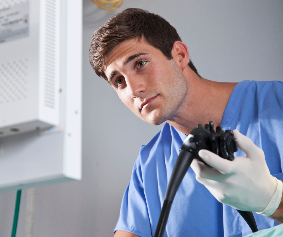
Push Enteroscopy Understanding Push Enteroscopy A focused procedure using a flexible tube to examine the small intestine for issues like bleeding or tumors. It provides a detailed view beyond traditional endoscopy. Introduction to Push Enteroscopy Push enteroscopy is a diagnostic procedure employed by gastroenterologists to examine the small intestine, a region not easily accessible through traditional endoscopy. This method plays a crucial role in identifying and diagnosing gastrointestinal issues such as bleeding, tumors, or inflammation within the small bowel. Indications for Push Enteroscopy Common reasons for recommending push enteroscopy include unexplained gastrointestinal bleeding, suspected small intestine abnormalities, or the need for tissue samples for further analysis. By directly visualizing the small intestine, gastroenterologists can provide accurate diagnoses and tailored treatment plans. During a push enteroscopy, a long, flexible tube with a light and camera at its tip, known as an enteroscope, is used to visualize the small intestine. This tube is gently guided through the upper gastrointestinal tract, enabling the physician to thoroughly inspect the lining of the small bowel. Patient Preparation for Push Enteroscopy To ensure a successful push enteroscopy, proper patient preparation is essential. Your healthcare provider will provide specific instructions, but typical preparations may include fasting for a certain period before the procedure. Clear communication with your healthcare team is crucial to understanding and following these guidelines accurately. Beyond physical preparation, it’s essential to mentally and emotionally prepare for the procedure. Feel free to discuss any concerns or anxieties with your healthcare team beforehand. Understanding the purpose and process of push enteroscopy can alleviate uncertainties and contribute to a smoother experience. In conclusion, push enteroscopy is a valuable tool in the diagnostic arsenal of gastroenterologists. By comprehending the procedure’s mechanics, indications, and adequately preparing for it, patients can actively participate in their healthcare journey, contributing to a more effective and efficient diagnostic process. Always consult with your healthcare provider for personalized advice tailored to your specific medical condition and needs. back to conditions page Facebook-f Youtube Linkedin-in Instagram
ERCP
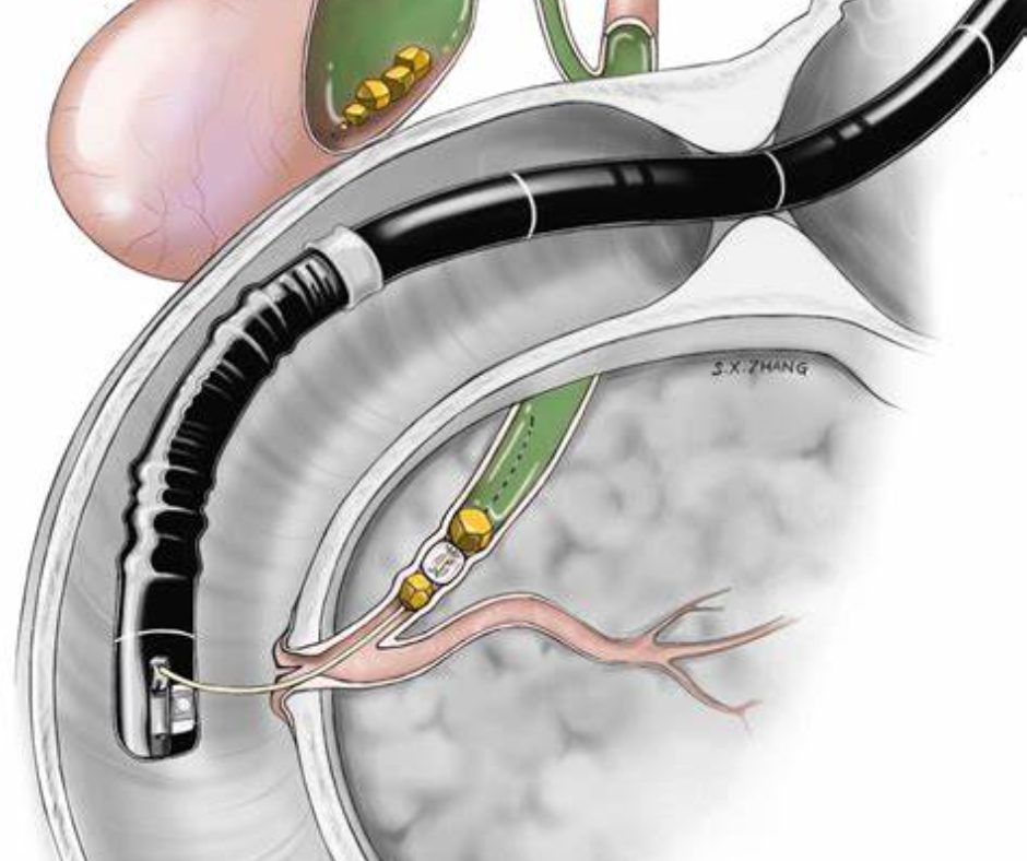
ERCP endoscopic retrograde cholangiopancreatography A diagnostic and therapeutic procedure employed by specialized gastroenterologists and surgeons to address issues related to the bile ducts, pancreatic ducts, and gallbladder. These vital anatomical structures play crucial roles in the drainage of bile and pancreatic juice, ultimately impacting digestive processes. Why is ERCP Performed? ERCP serves various purposes, with the most common being the identification and removal of gallstones obstructing the bile duct. Additionally, the procedure is utilized to investigate the causes of acute pancreatitis, alleviate duct blockages due to tumors, and treat leaks in the bile or pancreas ducts. Initial imaging tests, such as ultrasound, MRCP, or Endoscopic Ultrasound, usually precede ERCP to ensure a safer diagnostic approach. How is ERCP Performed? Conducted by trained gastroenterologists or surgeons, ERCP involves the use of an endoscope—a flexible tube with a camera and light. Administered under anesthesia, the endoscope traverses the mouth, esophagus, stomach, reaching the duodenum. The procedure allows for the introduction of wires, the application of treatments, and the injection of x-ray dye to visualize the ducts, facilitating decision-making on the course of action. How to Prepare for ERCP Preparation involves fasting for 6-8 hours before the procedure to ensure an empty stomach for optimal visibility. Discussion with your doctor about medications is essential, especially concerning blood-thinning agents and diabetes management. It’s crucial to disclose conditions like obstructive sleep apnea, lung issues, or heart conditions, as they may influence the anesthetic approach. An adult companion is required post-procedure due to the use of sedatives. What to Expect in the Endoscopy Suite Upon arrival at the endoscopy unit, a review of medical history, IV line insertion, and discussions with your doctor precede the procedure. You may need a breathing tube for general anesthesia. The ERCP room accommodates specific positioning for x-rays, and after the procedure, patients move to the recovery room for monitoring. Mild discomfort, sore throat, and grogginess are common post-ERCP experiences. Possible Complications of ERCP While generally safe, ERCP carries risks, including bleeding, infection, pancreatitis, perforation, aspiration, and reactions to anesthesia. Symptoms such as fever, severe abdominal pain, vomiting, or difficulty swallowing should prompt immediate communication with your doctor. Contact your doctor promptly if you experience symptoms such as fever, severe abdominal pain, vomiting, difficulty swallowing, abdominal bloating, bleeding, or any adverse reactions to medications. If biopsies were conducted, adhere to follow-up instructions to discuss the results. In conclusion, ERCP is a valuable tool in diagnosing and treating gastrointestinal issues, but awareness of potential complications and diligent post-procedural care are crucial for a successful outcome. Always stay informed and communicate openly with your healthcare team. back to conditions page Facebook-f Youtube Linkedin-in Instagram
Esophageal pH Study
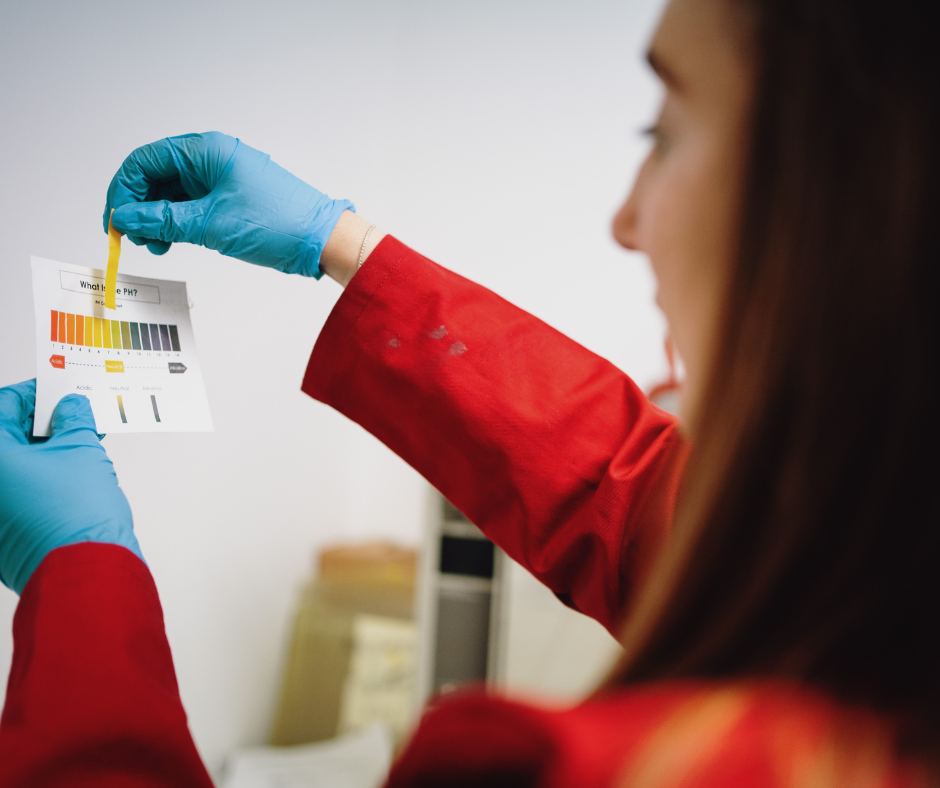
Esophageal pH Study exploring colon issues A diagnostic test that measures the acidity in your esophagus. It helps doctors understand if acid reflux or related issues are causing symptoms like heartburn. During the test, a thin tube is inserted through your nose into the esophagus to monitor acid levels over a period, providing valuable insights for diagnosis and treatment. The Significance of Esophageal pH Study Esophageal pH Study serves as a valuable tool in the realm of gastroenterology, allowing physicians to assess and diagnose conditions related to acidity levels in the esophagus. This procedure is commonly employed to investigate symptoms such as heartburn, regurgitation, and chest pain, providing essential insights into the underlying causes of these discomforts. Procedure Overview The Esophageal pH Study involves the insertion of a thin, flexible tube through the nose and into the esophagus. This tube is equipped with a pH sensor, which measures acid levels in the esophagus over a specific period. During this time, patients maintain their regular activities while the device records data, providing a comprehensive profile of acid reflux patterns. Patient Preparation Fasting Period: Patients are typically required to fast for a specific duration before the procedure, ensuring accurate readings. This involves abstaining from food and beverages, including water, for the designated timeframe specified by your healthcare provider. Medication Adjustments: Inform your healthcare provider about any medications you are currently taking, as certain drugs may interfere with the accuracy of the pH study. Adjustments or temporary discontinuation of specific medications may be necessary, and your physician will guide you through this process. Symptom Journal: Keeping a detailed journal of symptoms leading up to the procedure can assist your healthcare team in correlating pH data with specific events. Documenting the timing and intensity of symptoms provides valuable context for a more precise diagnosis. What to Expect During the Procedure On the day of the Esophageal pH Study, patients can expect a thorough explanation of the process by their healthcare provider. The insertion of the pH probe is a relatively quick and minimally invasive procedure. Throughout the monitoring period, patients are encouraged to resume their daily activities to ensure a comprehensive assessment of acid reflux patterns. In conclusion, the Esophageal pH Study is an invaluable diagnostic tool in gastroenterology, aiding in the identification and management of esophageal disorders. By understanding the significance of this procedure and adhering to the necessary preparations, patients contribute to the accuracy of the results, paving the way for more effective treatment strategies. If you have any further questions or concerns about the Esophageal pH Study, don’t hesitate to reach out to your healthcare provider. Your proactive involvement in the process is key to achieving the most accurate and beneficial outcomes from this diagnostic procedure. back to conditions page Facebook-f Youtube Linkedin-in Instagram
Flexible Sigmoidoscopy
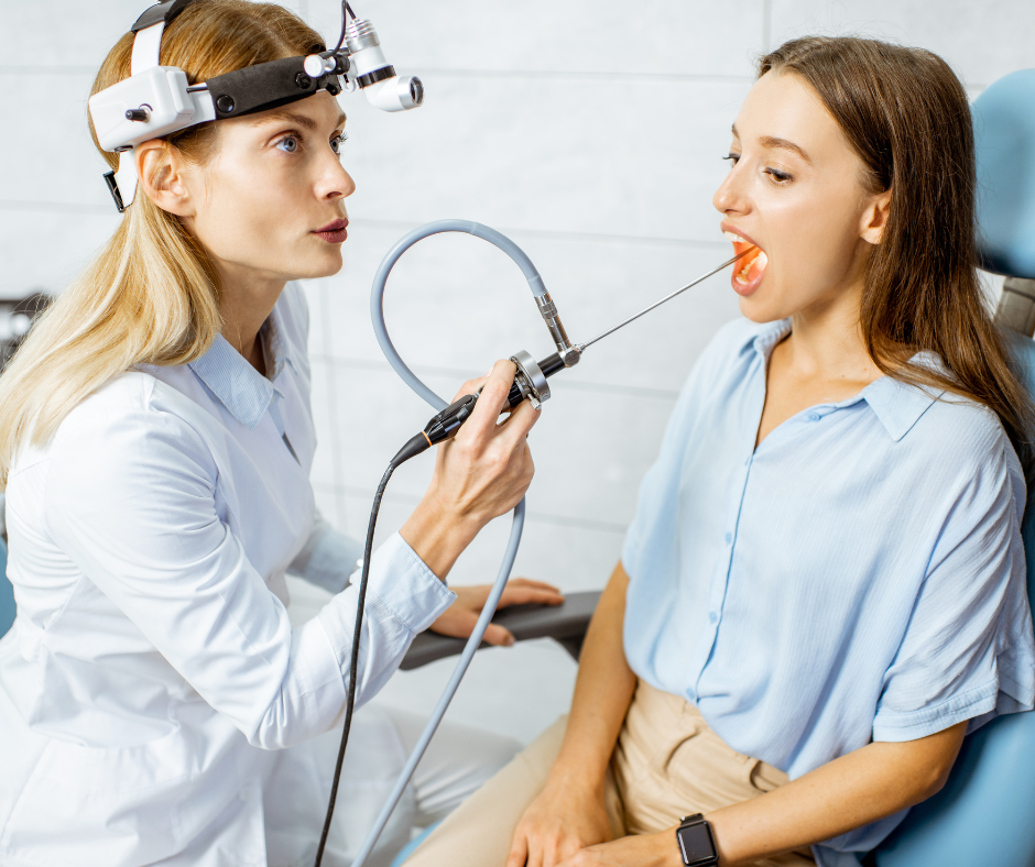
Flexible Sigmoidoscopy Understanding Flexible Sigmoidoscopy Flexible sigmoidoscopy is a diagnostic procedure commonly employed by gastroenterologists to examine the lower part of the colon, specifically the rectum and sigmoid colon. This minimally invasive test aids in the detection of various gastrointestinal conditions, such as colorectal polyps, inflammation, and other abnormalities. Purpose of Sigmoidoscopy The primary objective of flexible sigmoidoscopy is to assess and diagnose issues within the lower colon. This procedure allows gastroenterologists to inspect the lining of the rectum and sigmoid colon for abnormalities, including polyps and signs of inflammation. Detecting these issues early on is crucial for the prevention and management of colorectal conditions. Patient Preparation Guidelines To ensure the accuracy and effectiveness of the flexible sigmoidoscopy, proper patient preparation is imperative. Patients are advised to follow these guidelines: Bowel Preparation: A clear view of the colon is essential for accurate examination. Patients will be instructed to follow a specific bowel preparation regimen, typically involving the consumption of a laxative and a clear liquid diet. This step is crucial for the success of the procedure. Dietary Restrictions: Patients may need to adhere to certain dietary restrictions in the days leading up to the procedure. A gastroenterologist will provide detailed instructions tailored to individual health needs. Medication Adjustments: Inform your healthcare provider about any medications you are currently taking. Certain medications may need to be adjusted or temporarily discontinued to enhance the clarity of the examination. The Procedure During a flexible sigmoidoscopy, patients lie on their left side as the gastroenterologist gently advances a flexible, narrow tube with a light and camera (sigmoidoscope) through the rectum and into the sigmoid colon. This allows for a visual inspection of the colon’s interior. The procedure is relatively quick, typically lasting around 20 minutes. After the Procedure Following the flexible sigmoidoscopy, patients may experience some gas and bloating. It is essential to have someone accompany you, as the sedation used during the procedure may temporarily impair your coordination and judgment. The gastroenterologist will discuss the findings with you and provide any necessary recommendations for further evaluation or treatment. Flexible sigmoidoscopy is a valuable diagnostic tool in the realm of gastroenterology, offering a comprehensive examination of the lower colon. By adhering to the recommended preparation guidelines, patients can contribute to the success of the procedure and, ultimately, their overall gastrointestinal health. If you have any concerns or questions about flexible sigmoidoscopy, consult with your healthcare provider for personalized guidance. back to conditions page Facebook-f Youtube Linkedin-in Instagram
Colonoscopy
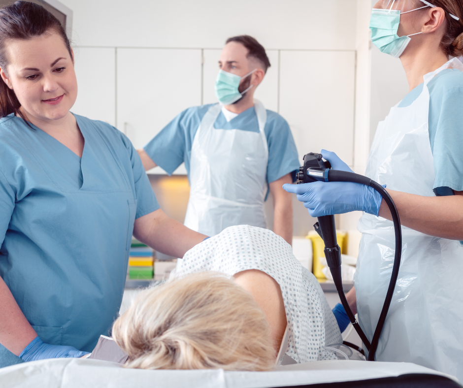
Colonoscopy exploring colon issues Essential for gastro health, it detects and treats various colon issues, from cancer to polyps, offering both diagnostic and therapeutic benefits, making it a crucial screening tool for colorectal health. Colonoscopy Overview Colonoscopy is a vital procedure conducted by gastroenterologists to directly visualize and examine the entire colon. This method plays a crucial role in diagnosing and evaluating various gastrointestinal disorders, such as colon polyps, colon cancer, diverticulosis, inflammatory bowel disease, bleeding, changes in bowel habits, abdominal pain, obstruction, and abnormal x-rays or CT scans. Moreover, it offers therapeutic benefits, allowing for the removal of polyps and control of bleeding. Addressing Common Concerns Despite its effectiveness, some individuals hesitate to undergo colonoscopy. Reasons range from lack of discussion with healthcare providers to concerns about the preparation process. While fear of the preparation is common, it is important to note that colonoscopy is typically not painful. Most procedures utilize “intravenous sedation” or “twilight sedation,” ensuring patient comfort and often resulting in amnesia regarding the procedure. For those without colorectal symptoms or family history, the recommended age for a first colonoscopy is 45, irrespective of gender. Family history influences the timing, with screening generally starting ten years earlier than the youngest family member diagnosed with colon cancer or polyps, or at age 40, whichever is younger. Individualized guidelines exist for rare syndromes. Alternative Screening Methods While alternative methods exist, colonoscopy remains the gold standard for accuracy in detecting colon cancer and polyps. Methods like flexible sigmoidoscopy or CT colography have their merits but may lack the diagnostic and therapeutic capabilities of colonoscopy. Bowel Prep: Overcoming a Common Obstacle The preparation process, often a concern for patients, can be tailored to individual preferences. Various methods, including liquid solutions, pills, and smaller volumes, ensure a clear colon for thorough examination. Consulting with a gastroenterologist helps determine the most suitable preparation method. Complications: Balancing Risks While complications are possible, they are rare and primarily associated with sedation administration. The benefits of colonoscopy in preventing colon cancer far outweigh the low risks, reinforcing its status as a safe and effective diagnostic and therapeutic procedure. Understanding one’s risk of developing colon cancer is essential. Factors such as family history, inherited disorders, obesity, smoking, inflammatory conditions, red meat consumption, and excessive alcohol intake contribute to overall risk. Timely discussions with healthcare providers can help assess individual risk profiles. In conclusion, colonoscopy stands as a cornerstone in the prevention and early detection of colon cancer. Understanding the procedure, addressing concerns, and prioritizing regular screenings are essential steps in maintaining gastrointestinal health. back to conditions page Facebook-f Youtube Linkedin-in Instagram