SBCE
SMALL BOWEL CAPSULE ENTEROSCOPY
A novel diagnostic tool that allows direct visualization of the small intestine. A vitamin-sized capsule containing a miniature camera is swallowed by the patient and takes thousands of pictures as it travels through the gastrointestinal tract.
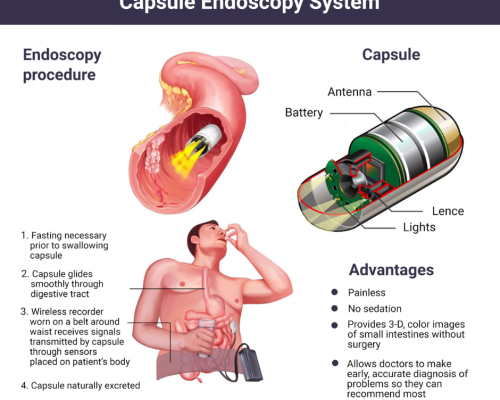
Overview of Small Bowel Capsule Enteroscopy
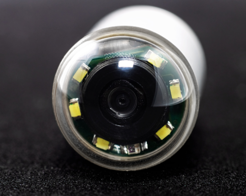
Small bowel capsule enteroscopy was approved by the FDA in 2001 as a tool to examine the small intestine, which previously could not be visualized well with traditional endoscopy. The capsule camera takes approximately 50,000 images during its travels through the GI tract, providing significantly more visualization than other methods. Capsule endoscopy is a non-invasive way to diagnose small bowel abnormalities and diseases, such as Crohn's disease, ulcers, celiac disease, small bowel tumors, and sources of GI bleeding.
Procedure
Patients fast overnight before capsule endoscopy. The next morning, they ingest the vitamin-sized capsule with water. Sensors are placed on the patient’s abdomen with adhesive pads, and are connected to a data recorder worn in a belt around their waist. Patients go about normal daily activities as the capsule camera takes images, while the sensors track its location and collect the images. After 8-12 hours, the sensors and recorder are removed, and the images are downloaded to a computer workstation for physician review. Patients pass the capsule naturally in their stool within 1-2 days.
Interpretation of Results
A physician carefully reviews the thousands of capsule images looking for abnormalities. Diagnostic findings include ulcers, bleeding, tumors, blockages, polyps, villous atrophy from celiac disease, and signs of Crohn’s disease. The images provide direct visualization to confirm diagnoses. However, capsule location can only be estimated based on image transit times. If an abnormality is seen, its exact location may be unclear. Another endoscopic procedure may be required to take biopsies or provide therapy.
Limitations
A major limitation of capsule endoscopy is the lack of therapeutic capabilities. It is strictly a diagnostic tool. Additionally, there is a small risk of capsule retention if a stricture is encountered. Capsule retention requires surgical or endoscopic removal. Some patients may have difficulty swallowing the capsule. Improper sensor placement or interference can also cause poor image quality. However, capsule endoscopy is considered very safe with few complications.
Small bowel capsule endoscopy allows detailed visualization of the small bowel mucosa. It is a minimally invasive and safe procedure to identify causes of GI bleeding, inflammation, tumors, celiac disease, and other small bowel abnormalities. Despite some limitations, capsule endoscopy is a revolutionary way to diagnose diseases of the previously hard-to-reach small intestine.